
|
|
Knee Surgery Review
|
|
Back in November I was playing indoor soccer as goalie in a pretty rough game and at one point I was driven into the ground pretty hard by this young bull of a guy. I made the save as I went down, and he helped me up and appologized for the roughness before he went upfield again, but my right knee was sore and immediately swelled and reddened. I didn't think anything of it, I get banged around a lot playing goalie and defense, especially in indoor soccer which is akin to soccer and hockey combined but without the pads. I noticed in the weeks after that the knee didn't have the strength it once had, and I found myself landing on my fanny sometimes when I tried to make very abrupt cuts or stops, but again didn't think much of it. In December I rented the arena for the girls team I coach to have a special practice and when a ball got stuck up in the nets I ran up to the wall and jumped up to grab the top of the glass wall to haul myself up and get the ball. When I launched off the ground my right knee made a pop noise and it felt like something moved inside it. I pulled myself up, got the ball, and dropped down on the left foot to prevent further shock to it. I immediately saw that I had damaged something inside the knee. It didn't hurt, but the leg didn't move normally, seeming to lag behind me as I tried to walk. The next morning the knee was swollen and very sore. Walking was difficult at best and I had to drag the leg around like a dead weight. I started the process of visiting my primary care physician after the holidays were over and I was sure the damage wouldn't heal itself. I found if my right foot slipped on the ice and twisted the knee I experienced excruciating pain, so I stopped doing that immediately. The dogs learned to quickly avoid my path when I walked through the room lest I growl at them to move, being so afraid I would trip over them and injure it worse. I made it through the "Oh you turned 40 since your last visit in 2000" visit to my wonderful doctor (truly wonderful) and he inspected the knee. Hearing the clicking noise it was making he sent me to the Orthopedic Doctor of my choice, who had repaired a teammates knee last year and received high marks from Joel (teammate) who is also a Doctor. The Orthopedic doctor checked me out on Tues (March 2) performing a McMurray Test on the knee and getting a positve result which indicated surgery necessary to fix the problem. I had described the symptom as feeling like a piece of cartelage had broken loose and slipped occasionally if weight was placed on the leg from certain angles. Also, the stability of the knee seemed compromised. He scheduled Orthoscopic Surgery for the next day (!!!) and I returned Weds. morning in my jammies, changed into the dreaded gown and slippies, and got comfortable on a gurney while they hooked me up to various devices and comforted me with small talk. The Anestheiologist came in and started me on the General Anesthesia saying to count up to 10 and not be concerned about reaching it as I probably wouldn't. I definately remember hitting 10 but nothing after that, though the nurses told me later I was at 13 when the lights went out.
|
 The knee joint is made up of three bones and a variety of ligaments. The knee is formed by the femur (the thigh bone), the tibia (the shin bone), and the patella (the kneecap). Several muscles and ligaments control the motion of the knee and protect it from damage at the same time. Two ligaments on either side of the knee, called the medial and lateral collateral ligaments, stabilize the knee from side-to-side.
 The anterior cruciate ligament (ACL) is one of a pair of ligaments in the center of the knee joint that form a cross, and this is where the name "cruciate" comes from. There is both an anterior cruciate ligament (ACL) and a posterior cruciate ligament (PCL). Both of these ligaments function to stabilize the knee from front-to-back during normal and athletic activities. The ligaments of the knee make sure that the weight that is transmitted through the knee joint is centered within the joint minimizing the amount of wear and tear on the cartilage inside the knee. The weight-bearing surfaces of your knees are covered with a layer of cartilage (referred to by doctors as"articular cartilage"). There are also two shock absorbers in your knee on either side of the joint between the cartilage surfaces of the femur and the tibia. These two structures are called the medial meniscus and the lateral meniscus. The menisci are horseshoe-shaped shock absorbers that help to both center the knee joint during activity and to minimize the amount of stress on the articular cartilage.
|
|
|
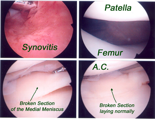
Making three incisions (one on each side and one above) the Surgeon first discovered a large amount of Synovitus or inflamed tissue in the Synovial region of the knee. Synovitis is the inflammation of a synovial (joint-lining) membrane, usually painful, particularly on motion, and characterized by swelling, due to effusion (fluid collection) in a synovial sac. Synovitis is a major problem in rheumatoid arthritis, in juvenile arthritis, in lupus, and in psoriatic arthritis. He removed the inflamed tissue Orthoscopically and continued on his investigation. In the area of the Medial Meniscus he found my little slipping piece of knee. It was a broken section of the Medial Meniscus shown in the above set of photos (Bottom Left) lifted up with the probe and (Bottom Right) resting as he found it.
|

|
|
He must have opened up one of the incisions a bit larger because the surgeon was able to remove the broken section of Meniscus intact. The set above shows the broken section (note the jagged edges) being lifted out and away. After removal he inspects my ACL (Anterior Cruciate Ligament) and my LM (Lateral Meniscus) for damage but they are both healthy.
|

|
|
All that now remained was what the surgeon calls "housekeeping". The third set of photos above shows roughness in the cartilage left after removal of the broken section. Orthoscopically this is ground off and returned to smoothness. Before closing he inspects the attachment end (opposite end view) of the ACL and finds it healthy. Finally the lower right photo in the 3rd set shows the offensive bit of Meniscus that caused all this ado.
|
|
The evening of the surgery was painful. My family did a wonderful job of making me comfortable, setting up a nice bed desk for my laptop, bringing me water with lemon. It was like being royalty. Three big Vicodins banished the pain to the outher regions and I had a pretty easy night. Yesterday I cut my pain med dosage from 2 to 1 pill every 4 hours and started using the leg a bit during restroom visits. Today was easier and I went to the doctors office for the followup without the crutches. He seemed pleased by that and said I should expect a full recovery. Ice 6 to 8 times per day for the swelling and more rest this weekend, then PT starting next week. It wasn't as bad as I feared it might be, and I am left with an even greater appreciation of good doctors, nurses, and modern medicine in general. Greg
|
|
Home | Gregs Page | Lori's Page | Our House | News | Genealogy | Logans Page | Lindens Page | Alfalfa Hay | Photos | Soccer | Classroom | Top Copyright 2001 Last Updated 03/05/04 Powered by Arachne |